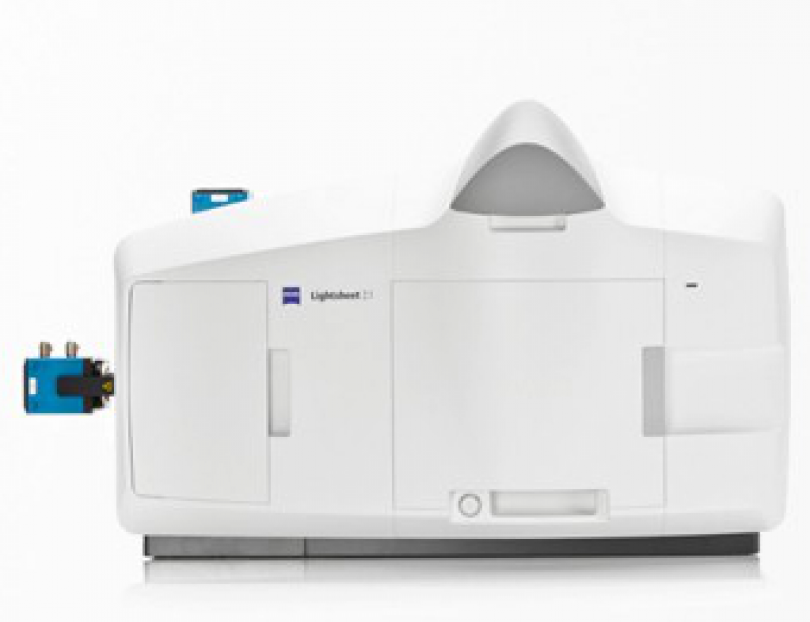
Zeiss Lightsheet Z.1

lightsheet Z1
Lightsheet microscopy is a fluorescence-based imaging technique that is growing in popularity. It outperforms other techniques in its ability to rapidly capture large optically sectioned three dimensional datasets, whilst keeping photo-toxicity and bleaching to a minimum. It therefore lends itself well to imaging samples larger than those typically attempted by conventional fluorescence microscopy such as tissues, whole organs and even embryo development.
Microscope body:
Custom
Motorised Stage:
Yes
Temp Control:
Heated Chamber
CO2:
Stage Insert
Software:
Zen


Common Applications


Lightsheet microscopy Lighsheet microscopy creates and positions a sheet of excitation light across the focal plane of a collection objective. In doing so it restricts excitation events in a particular plane of the sample resulting in an optically sectioned image drastically reducing light emanating from outside of the focal plane. Moving the sample through this plane allows rapid collection of sectioned 3D data.
Tissue Clearing: Tissue clearing is a preparation technique with the aim of reducing the inherinatly scattering nature of tissue. There are several means by which this is achieved but all enable clearer and deeper penetration into the sample using light microscopy.
Live Cell Imaging Lightsheet imaging can also be done in brightfield and/or fluorescence on a small live and fairly transparent specimen (e.g. Spheroids, Organoids, zebra fish …). The chamber can be heated to the temperature required and the specimen is immersed in its medium during the imaging. 


Objective Lenses

Collection Objectives

MAG N.A CORRECTIONS IMMERSION MISC.
2.5C Clearing 0.12 Fluar Air nd: 1

5x

0.16 Plan Neofluar Air nd = 1.33-1.56
10X 0.5 Plan Apochromat Water nd = 1.336
20x 1 Plan Apochromat water nd: 1.32 -1.4
20x Clearning 1 Plan Neofluar Clearing nd = 1.42-1.45
40x 1 Plan Apochromat water VIS-NIR


Illumination Objectives

MAG N.A CORRECTIONS IMMERSION MISC.
LSFM 5x 0.1   Air  

LSFM 10X

0.2   Air  
5x Focusing 0.1 Medium Refractive Index 1.33 -1.58 Air  

Light Sources

Laser lines: 405 nm, 445 nm, 488 nm, 515 nm, 561 nm, 638 nm 

Filters

The system can hold one of two filter wheels positined just before the two cameras. Note these filters are not to separate the excitation and emission lightpath (as these are already separate), these filters are designed to split the emitted light to separate cameras for simultaneous imaging. 


Camera Filter Wheel 1 

NAME 

Camera 1 

DICRHOIC 

Camera 2 

Universal Blue-Green 

SP 490 

BS 490 

LP 505 

CFP-mCherry 

BP 460-500 

BS 510 

LP 585 

GFP-RFP 

BP 505-545 

BS 560 

BP 575-615 

YFP-RFP 

BP 525-545 

BS 560 

BP 575-615 

CY3-DRAQ5 

BP 575-615 

BS 640 

LP 600 


Camera Filter Wheel 2 

NAME 

CAMERA 1 

DICRHOIC 

CAMERA 2 

DAPI-CY3 

BP 420-470 

BS 510 

BP 575-615 

CFP-YFP 

BP 460-500 

BS 510 

BP 525-565 

GFP-RFP 

BP 505-545 

BS 560 

BP 575-615 

GFP-DRAQ5 

BP 505-545 

BS 560 

LP 660 

GFP-mCherry 

BP 505-530 

BS 560 

LP 585 


Detectors

This instrument is equipped with 2 PCO Edge 5.5 cameras. They are configured to allow simultaneous imaging of two channels