Our state-of-the-art facility welcomes researchers, students, and science enthusiasts to explore the unseen with cutting-edge technology. Here, we offer a range of advanced microscopy services designed to push the boundaries of imaging and analysis.
40+ Microscopes
400+ Users/Year.
Flat fee options
In-depth Training
How do I gain access?
Talk to us about your research questions.
Registering on our ACLS booking system marks the start of your onboarding process. For more information on how to register, please visit our new user page.
Should you have any questions about our microscopes, their capabilities, or how they can assist your research, feel free to reach out for guidance. You can discuss your needs with a microscopy expert by sending an email to kglmf@unsw.edu.au.
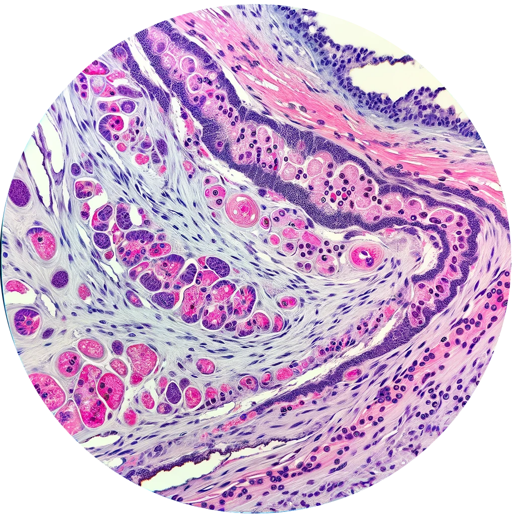
Brightfield Microscopy
Brightfield Microscopy offers a simple yet powerful way to view stained or pigmented specimens with high contrast. Ideal for biological research and medical diagnostics, our service provides clear, detailed imaging and expert support.
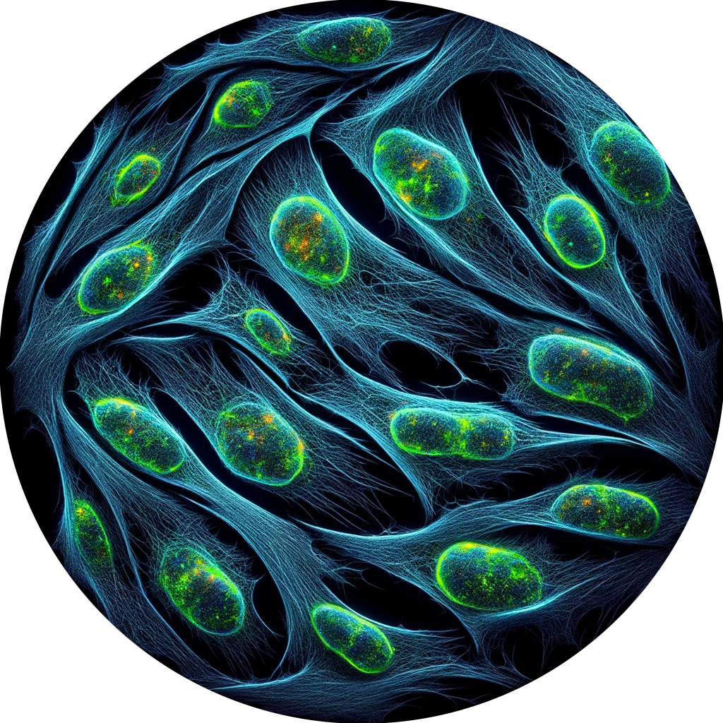
Fluorescence Microscopy
Fluorescence microscopy uses fluorescent dyes to illuminate and study specific components within cells and tissues. It excites these dyes with specific wavelengths of light, causing them to emit light that forms detailed images. This technique is crucial for high-resolution, targeted observations in biological and biochemical research.
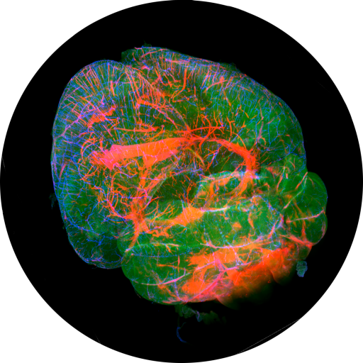
Optical Sectioning techniques for improved 3D visualisation
We offer a range of optical sectioning methods, including confocal, lightsheet, spinning disk confocal, and apotome, each providing unique, high-resolution 3D imaging for diverse research needs.
Fluorescence Lifetime Imaging Microscopy
Fluorescence Lifetime Imaging Microscopy (FLIM) provides a dynamic view of cellular processes by measuring the decay rate of fluorescence, offering a window into the molecular environment of biological samples. Unaffected by dye concentration or photobleaching, FLIM excels in live-cell imaging for real-time, quantitative analysis. This advanced microscopy technique is crucial for cutting-edge research in cell metabolism, protein interactions, and tissue diagnostics.
Super-Resolution
Embrace the power of super-resolution at our facility, where we offer advanced techniques like STORM, PALM, STED, SIM, and Airy-scan. These cutting-edge methods break through the traditional limits of optical microscopy, allowing you to observe details at the nanoscale. Whether you're investigating intricate cellular structures or dynamic molecular processes, our super-resolution technologies provide unparalleled clarity and detail.
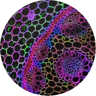
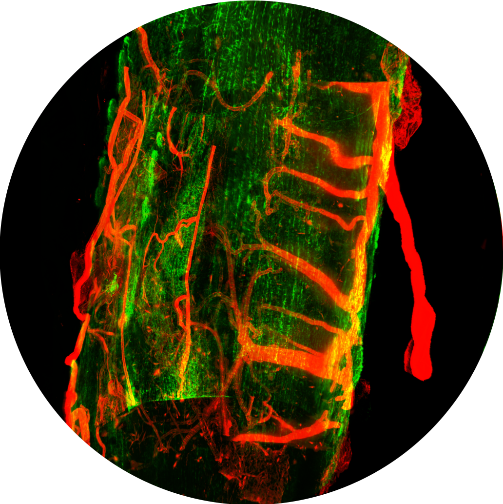
Multi-photon imaging for deeper tissue imaging
Our Multi-Photon equipped microscope offers a remarkable increase in penetration depth for deep tissue imaging. This advanced technique excels in visualizing thick biological samples, extending far beyond the capabilities of conventional microscopy. Additionally, it is adept at Second Harmonic Generation (SHG) imaging, enabling the detailed observation of non-fluorescent structures like collagen.