Instruments

Searching all Light Microscopy Facility instruments. Search all instruments.
3i Lattice Lightsheet

Lightsheet

The 3i Lattice LightSheet allows for gentle 4D imaging of live cells. The system creates an ultra-thin Gauss-Bessel light sheet that can be used to image cells for short or long time periods. This system has high spatial and temporal resolution due to the unique lattice lightsheet. Images can be captured in excess of 500 frames per second. 

3i VIVO™ Microscope

Fluorescence Widefield Spinning Disk Confocal Intravital

The 3i VIVO microscope is ideally suited for intravital imaging. Its large stable stage can easily accommodate sizable tissue samples along with clamps and manipulators to aid stabilization. Equipped with a spinning disk unit for high speed optical sectioning, paired with an image intensifier, this system can achieve very high frame rates making the most of every photon. Additionally, a pulsed 532nm ablation laser is available for targeted tissue damage. 

Aperio XT Slide Scanner

The Aperio ScanScope XT scanner is a brightfield microscope that is used to digitalise traditional histochemicaly stained tissues mounted on a microscope slides. You can choose between 20X and 40X magnification and scan any required size of the tissue. The digital slides are viewed and analysed using Aperio software applications (ImageScope). The image format is .svs which is a form of a tiff file, that can be converted into JPEG2000 for faster processing and presentation. The Aperio XT scanner can autoload up to 120 glass slides (1x3") as well as manually scan your slides one by one if required.

Bruker Multimode 8

AFM

The Bruker MultiMode® 8 (MM8) is an advanced atomic force microscope (AFM) and Scanning Tunnelling Microscope (STM). Its PeakForce Tapping technology enables exceedingly low tip-sample force interactions compared to tapping mode, making it particularly useful for delicate samples by allowing the user to control the maximal force imparted to the sample. The MM8 also features the Bruker ScanAsyst® software, automating the optimisation of scanning parameters for users to obtain quality images.

Chemicon Advanced Tissue Arrayer

Specimen Preperation

Chemicon Advanced Tissue Arrayer ATA100 allows you to create customized arrays of multiple tissue specimens on a single histologic slide. This instrument allows to selectively remove core tissue samples from paraffin blocks and arrange those tissue samples into an array within a recipient paraffin block. The newly formed tissue array paraffin block is then ready for further histology technique processing.

EasyGel And SmartClear II

Clearing Specimen Preperation

EasyGel is a system designed for speeding the process of producing tissue-gel-hybridization samples. The SmartClear II system is a commercial instrument that allow fast, easy and reliable tissue clearing of tissue-gel-hybridization samples. The system uses stochastic electrotransport technology to remove lipids from tissue-gel-hybridization samples, thereby producing optically transparent samples. Samples that have been optically cleared with the SmartClear II system can be imaged by a number of light microscopy techniques such as confocal and lightsheet. 

FluoView FV1200

Fluorescence Confocal DIC

The FV1200 is a filter based upright confocal system. A workhorse of our confocal systems it is well equipped for standard fixed samples where high resolution and sensitivity are required for localization based studies. Additionally as one of the few upright confocal systems within the BMIF it provides a means for visualizing water submerged opaque samples.

Image Analysis Computers

Image Analysis

We currently have 8 image analysis computers, all of them accessible remotely, whilst 4 are also accessible locally with 4k 48inch displays so you can see your data in its best light. We also have a varied range of commercial software packages available on many of these computers

IncuCyte S3

Fluorescence Phase Contrast Widefield High Throughput

he IncuCyte S3 is a widefield inverted live cell system engineered with long term high throughput imaging in mind. Housed inside a tissue culture incubator, the imaging module maintains the most stable imaging conditions possible ( 37C temperature, humidity and 5% CO2). The system comes with a selection of versatile inserts allowing you to image your cells in a range of vessels.

IncuCyte SX5

Fluorescence Phase Contrast Widefield High Throughput

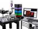
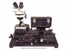
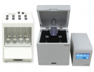
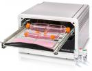
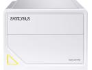
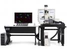
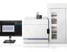
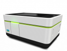
The Incucyte SX5 is engineered for long-term, high throughput, widefield microscopy. The system is compatible with a range of plates and cell culture flasks in stable conditions (37°C temperature, stable humidity and 5% CO2), with automated imaging for long-term experiments.  

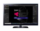
Images can be acquired over time and analysed in real time. The SX5 supports a wider range of fluorescent imaging than the S3, with up to five fluorescent channels available. The acquisition software is loaded with a variety of analysis tools and modules, details below.  

JPK Nanowizard 4 XP with Hybrid stage

AFM

The NanoWizard® 4 XP atomic force microscope is an advanced Atomic Force Microscope (AFM). AFM is a very high-resolution type of Scanning probe microscopy (SPM) which acquires data by “feeling” or “touching” the surface of a sample with a mechanical probe


Leica Bond Rx

Specimen Preparation OPAL

The Bond RXm automated staining system increases reproducibility of IHC (chromogenic or immunofluorescent) and multiplex staining. Custom protocols can be programmed for projects, complete with temperature control for steps such as antigen retrieval or TSA staining. Long staining protocols, such as OPAL multiplex can be set to be completed as an overnight run, while shorter protocols can be run during the business day

Leica Bond Rx (UNSW & Ramaciotti)

Specimen Preperation

The BOND RX provides an easy way to automate the preparation of samples. In combination with PerkinElmer Opal™ Kit it is also useful for multiplexing allowing up to 9 fluorescent labels within a single sample.

Leica DM IL

Fluorescence Widefield

The Leica DM IL microscope is used to illuminate whole cells; observing and analysing a cell as a single entity. The DM IL is an inverted epifluorescence microscope, with transmitted light LED back illumination and an EL6000 Fluoro system.

Leica RM2135 Rotary Microtome

Specimen Preperation

Leica RM2135 rotary microtome is used to cut very thin cross-sections of specimens (usually 4 µm) for routine and research histology studies in the fields of biology and medicine.  This instrument is designed for cutting soft and harder paraffin embedded specimens, as long as they are suitable for being cut manually.  The sectioning thickness setting range can be from 1 µm to 60 µm.  High quality paraffin sections could be used for varying staining, such as Haematoxylin and Eosin stain (H&E), immunohistochemistry (IHC), Immunofluorescent staining (IF) and In situ hybridization (ISH).

Leica SP8 DLS

Confocal Lightsheet

The Leica TCS SP8 DLS (Digital LightSheet) is an inverted laser scanning microscope capable to be used for confocal imaging and light sheet imaging. This microscope is able to image multi-colour, live, fixed or aqueous-based cleared specimens utilising a combinations of 405, 458, 488, 514, 561 and 635 nm excitation lasers through the employment of the AOBS with spectral high quantum efficiency detectors. The advantage of the two combined features (confocal/Lightsheet) on a single equipment allow to perform, with the confocal module, local photo activation or photo bleaching where the resulting dynamics can be capture via lightsheet imaging on thick sample (up to 2 mm thick and 1 cm long).

Nikon A1 Intravital

Confocal Intravital

The Nikon A1 intravital microscope is an inverted confocal system designed for fast stable imaging over time. With two scanners, a standard Galvano scanner for achieving images with a high signal-to-noise ratio and a resonant scanner for when speed is of upmost importance, this system is suitable for a wide range of experiments. Equipped with 4 dedicated PMTs for reflective/fluorescence imaging, 2 of which as high efficiency GaAsP detectors, and a hardware solution (Perfect Focus System) for preventing drift during imaging this system yields both high sensitiy with speed and stability.

Nikon AXR

Confocal Resonant Scanner Automation

The Nikon AX-R2 is a confocal system with 2k x 2k resonant scanning and 8k x 8k Galvano scanning technologies. The system is designed to balance sensitivity and resolution, with the capacity to perform spectral scanning and unmixing. The system is fitted with 4 detectors and 404/488/561/660nm lasers for a wide range of applications.

Nikon N-Storm (Ramaciotti)

Super-resolution TIRF Widefield Funded by Ramaciotti Foundation

This is a total internal fluorescence (TIRF) microscope built on an inverted Nikon Ti2-E microscopeThe TIRF modality allows user to image molecules at surface of cells and apply super- resolution imaging techniques such as PALM and STORM. It is equipped with piezo Z stage and Nikon Perfect Focus System 4, making it ideal for long term imaging. It is equipped with stage top incubator and objective heater as well as CO2, allowing live cell imaging. With quad filter, 4 solid state laser lines,

Nikon Ti2 (Biosciences)

Fluorescence Widefield Phase Contrast DIC Calcium Imaging

The Nikon Eclipse Ti2-E is an inverted widefield system offering a large field of view, high-quality optics and an industry-leading focus stabilisation system (Nikon Perfect Focus System). he system software includes Nikon JOBS, allowing automation of experimental protocols, including on the go analysis and cell identification. Additionally, a CoolLED pE-340Fura light source enables rapid ratiometric imaging of rapid calcium dynamics.

Nikon Ti2 Spinning Disk (Ramaciotti)

Spinning Disk Confocal Fluorescence Widefield High Throughput Phase Contrast DIC Funded by Ramaciotti Foundation

The Nikon Eclipse Ti2-E is an inverted widefield system offering a large field of view, high-quality optics and an industry-leading focus stabilisation system (Nikon Perfect Focus System). With a stage insert providing temperature and CO2 control, this system is ideal for live-cell imaging. The system software includes Nikon JOBS, allowing automation of experimental protocols, including on the go analysis and cell identification. In addition to these features, this microscope is connected to a Cetoni liquid-handling unit, allowing for automated liquid exchange. Whether you are performing live-cell experiments, sample screening or automated experimental protocols, this system can perform the task with minimal researcher input.

Nikon TIRF/HiLo

Florescence TIRF HiLo Intravital Widefield

This microscope combines a total internal fluorescence (TIRF) and highly inclined and laminated optical sheet modalities in one instrument. TIRF modality allows user to image molecules at the surface of cells and apply super- resolution imaging techniques such as PALM and STORM. HiLo modality offers the possibility of exciting a thin inclined plane within cells or thin layer of cells within few micrometers of a live animal tissue. Automatized variability of angle of HiLo emergence and camera imaging allows for fast sectioning of sample.

Olympus Slideview VS200

Slide Scanning Fluorescence Brightfield Dark field Polarization

The Olympus SLIDEVIEW VS200 is a high throughput bright-field and fluorescent slide scanner that facilitates digitalisation of archival, diagnostics, teaching and research histology and cytology microscopy slides. 
This system offers users the choice of five imaging modes: Brightfield, Darkfield, Phase Contrast, Polarization and Fluorescence.

Operetta CLS

Fluorescence High Throughput Spinning Disk Confocal

The Operetta CLS is a multi-modal high-content microscopy system. Designed for high throughput, it can capture both widefield and spinning disk confocal images. Setup a single well in a 96 well plate and simply allow the system to replicate those settings across the entire plate. With an automated water immersion objective you also don’t have to sacrifice resolution for high throughput multipoint positioning.

Phasefocus LiveCyte

Ptychography

The Phasefocus Livecyte is an inverted system for live cell imaging. This microscope has Quantitative Phase Imaging (QPI, also known as Ptychography) a label-free approach that utilises relative phase shift information to produces images of cells and tissues with high contrast and high fidelity with low phototoxicity and photobleaching. The system is also equipped with widefield fluorescence capabilities for researchers that wishes to identify cells and cellular components with high specificity.