The MRI Facility at UNSW
The MRI facility at the UNSW’s BRIL is one of the most powerful small-animal MRIs in Australia and is able to provide users with state-of-the-art small animal and sample imaging, and MRI biomarker assessment. As part of the National Imaging Facility the scanner can be accessed by researchers from institutions from all over the country.
With a magnetic field of 9.4T and a bore of 20cm it is ideally suited for high resolution imaging and biomarker assessment from a wide variety of samples and tissues. Currently the facility is able accommodate samples and animals of sizes up ~80mm in diameter and depending on measurement time, can yield anatomical information with an isotropic spatial resolution of up to 30-40µm. For an overview of MRI applications and references please see the techniques page.
The facility is equipped with a full featured animal anaesthesia and monitoring system, enabling measurements on all kinds of small live animals including rodents (mice and rats). As the MRI scanner is part of a PC2 certified laboratory and animal facility, investigations can be carried out on animals requiring this level of containment.
MRI Facility Technical Data
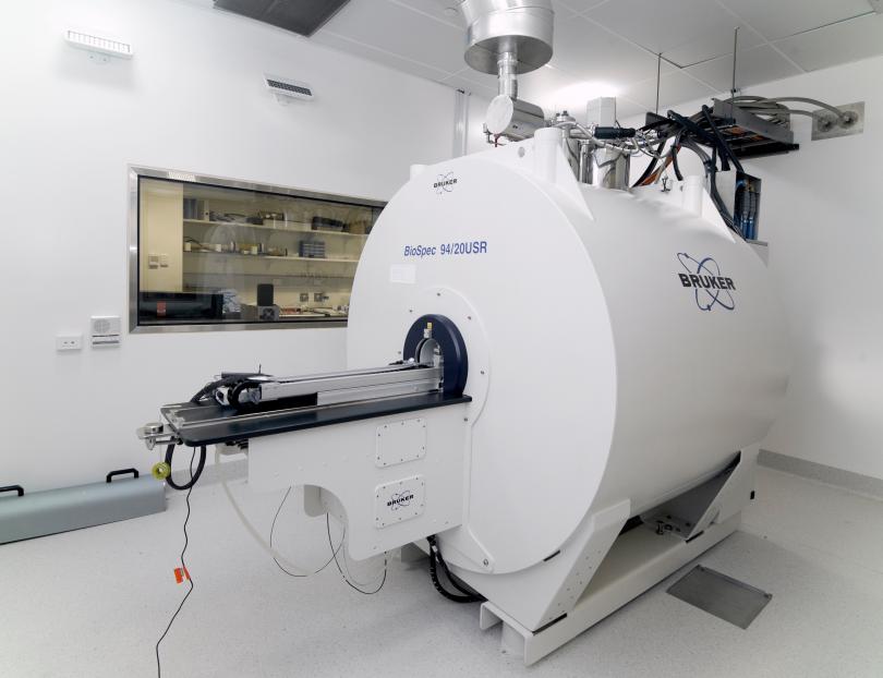
System Name: Bruker BioSpec Avance III 94/20
Magnet:
· cryo-cooled, actively shielded, zero-boil-off magnet
· Field strength: 9.4T
· Resonance frequencies: H: 400MHz
· Bore diameter: 20cm
· Field homogeneity DSV: 120mm
· Field homogeneity peak-peak: 10ppm
Gradient System:
The facility has two sets of gradients: Depending on the application the standard high power gradient system an additional Micro imaging gradient insert can be chosen. The micro imaging system provides substantially higher gradient power, with the draw-back of smaller inner diameter, making it especially suitable for micro-imaging applications and physiological measurements requiring high gradient strength, such as diffusion imaging.
- High Power Gradient System wide bore:
Inner diameter: 114mm
Max. Gradient strength: 660 mT/m
Max linear slew rate: 4570 T/m/s
- High Power Gradient Insert for Micro-Imaging:
Inner diameter: 60mm
Max. Gradient strength: 1000 mT/m
Max linear slew rate: 11250 T/m/s
RF-System:
Transmitter Channels: 2x1kW
Receiver Channels: 1 Broadband channel (capable for H and X-nuclei), 8 parallel proton-receiver channels
Software:
The scanner software is based on the Bruker TopSpin package that also controls most Bruker NMR scanners for chemical applications. On this platform the ParaVision 5.1 software package supports all imaging applications.
The software available at BRIL includes the following special application and data evaluation packages:
· Spectroscopy Package
· Diffusion Tensor Imaging Package
· SWI Package
· Spiral Imaging Package
· Intragate Cardiac Imaging Package
· Ultrashort Echotime Imaging (UTE)
Apart from the standard image acquisition packages, in-house built pulse sequences can be implemented or imported from other Bruker scanners for special applications. Currently BRIL provides in-house MRI pulse sequences for Oscillating gradient diffusion imaging applications and CEST imaging.
RF coils
Cryo-cooled RF coils
The facility is equipped with a 20mm Cryo-probe which can be installed for certain mouse and sample experiments to provide very high SNR for high-resolution imaging and spectroscopy studies.
Standard RF-coils
The facility has a variety of coils available to suit a wide range of proton imaging and spectroscopy applications. The currently available proton coils are listed below:
|
Coil Type |
Diameter |
Applications |
|---|---|---|
|
Volume Transmit Resonator |
86mm |
General transmission for surface coil operation and imaging of larger samples and animals (e.g. obesity studies) |
|
Volume Transmit/Receive Resonator |
72mm |
Homogeneous volume imaging of large to medium size samples and animals mainly rats, fits the rat and large mouse anaesthesia bed |
|
Volume Transmit/Receive Resonator |
50mm |
Homogeneous volume imaging of medium size samples and mice as well as smaller rats, fits large mouse anaesthesia bed |
|
Volume Transmit/Receive Resonator |
35mm |
Homogeneous volume imaging of medium size samples and mice as well as smaller rats, fits large mouse anaesthesia bed |
|
Mouse Head Volume Transmit/Receive Resonator |
23mm |
Dedicated volume coil for homogeneous High SNR imaging of mouse brains. With the in-house built sample holder this can also be used for samples up to 20mm diameter |
|
Volume Transmit/Receive Resonator |
15mm |
Homogeneous, high SNR volume imaging of small samples anaesthesia bed |
|
Surface Receive-only Coil |
20mm (surf) |
High SNR imaging of localised animal body regions or small samples. Coil must be operated with the 86mm Transmit Resonator. Coil delivers high SNR but an inhomogeneous signal over the FoV. |
|
Surface Receive-only Coil |
10mm (surf) |
High SNR imaging of localised animal body regions or small samples. Coil must be operated with the 86mm Transmit Resonator. Coil delivers high SNR but an inhomogeneous signal over the FoV. |
X-nucleus RF-coils
The facility has a variety of coils for non-proton imaging and double resonant 1H/x-nucleus imaging and spectroscopy. The supported nuclei can be found in the list below
|
Nucleus / Coil Type |
Diameter |
Example applications |
|---|---|---|
|
13C/1H Double resonant Surface Transmit-Receive Coil |
20mm (surf) |
Metabolic 13C Spectroscopy and imaging studies using glucose or similar tracers |
|
19F/1H Double resonant Surface Transmit-Receive Coil |
20mm (surf) |
Oxygenation assessment, cell tracking, targeted imaging |
|
31P/1H Double resonant Surface Transmit-Receive Coil |
20mm (surf) |
Metabolic 31P studies (ADP, etc.) |
|
129Xe/1H Double resonant Linearly Polarized Volume Transmit-Receive Coil |
35mm |
Lung imaging, ventilation imaging |
Animal Handling and Monitoring System
The facility is located inside a fully equipped PC2 animal laboratory, capable of handling animals and samples requiring this containment. The following list gives an overview over the available equipment:
· Automatic sample/animal positioning system
· Animal beds for both rats and mice
· Animal bed heating facility + additional heating blankets
· Built in isofluorane gas anaesthesia station
· Animal Monitoring unit:
- Breathing monitor/triggering
- Temperature monitor
- ECG monitoring/triggering
Please see the techniques page for more information