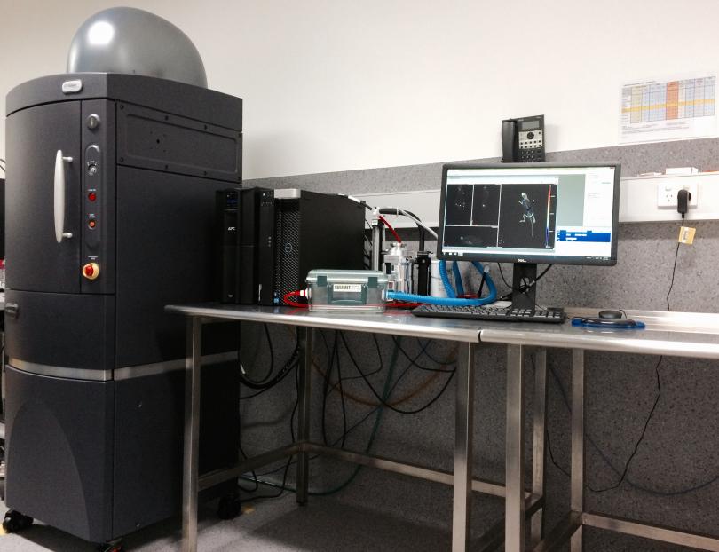
IVIS® SpectrumCT
The IVIS® SpectrumCT pre-clinical in vivo imaging system provides a highly sensitive means to image bioluminesence and/or fluorescent reporters in vivo, ex vivo and in vitro. Bioluminescence imaging (BLI) of transgenic animals or cells expressing luciferase requires the administration of luciferin bioluminescent substrate prior to imaging. In a typical luciferase imaging setting, imaging of the animals takes between 10-20 minutes for each set, however this can increase to 45 minutes depending on the model.
The system is integrated with low dose microCT to support longitudinal imaging. The IVIS SpectrumCT enables simultaneous molecular and anatomical longitudinal studies, providing researchers with essential insights into complex biological systems in small animal models. The constant horizontal gantry motion and the flat panel detector provide low-dose imaging and automated optical and microCT integration. The stable revolving animal platform table rotates 360° to acquire full 3D data. Multiple animals can be scanned simultaneously while maintaining an average dose per scan at about 13mGy, with a scanning and reconstruction time of less than a minute. Optical and microCT modalities can also operate independently. Multispectral fluorescence imaging (400-900nm wavelength) can be performed with the system for 2D or 3D imaging. Deep tissue detection and quantification can be achieved through trans-illumination available on this system. The system is fully equipped with its own isoflurane anaesthetic system and is capable of imaging up to five mice simultaneously.
LivingImage Software
Data acquisition and analysis are performed using the LivingImage software. The software allows the data to be reviewed individual and regions of interests quantified as the data acquired from the IVIS systems can be expressed in radians (photons/sec/cm2/sr) for luminescence and radiant efficiency for fluorescence, providing a quantitative means for comparing data between animals.
Please see the techniques page for more information