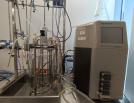
Spinning Disk Confocal Fluorescence Widefield High Throughput Phase Contrast DIC Funded by Ramaciotti Foundation
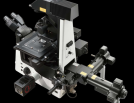
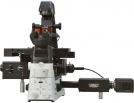
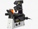
The Nikon Eclipse Ti2-E is an inverted widefield system offering a large field of view, high-quality optics and an industry-leading focus stabilisation system (Nikon Perfect Focus System). With a stage insert providing temperature and CO2 control, this system is ideal for live-cell imaging. The system software includes Nikon JOBS, allowing automation of experimental protocols, including on the go analysis and cell identification. In addition to these features, this microscope is connected to a Cetoni liquid-handling unit, allowing for automated liquid exchange. Whether you are performing live-cell experiments, sample screening or automated experimental protocols, this system can perform the task with minimal researcher input.