
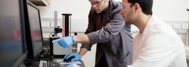
Tyree X-ray micro-CT Facility
- Link to the School of Minerals and Energy Resources Engineering in Tyree can be accessed here.
- The Pricing for access to Tyree x-ray CT facility can be found here.
- Take a virtual tour of the Tyree X-ray micro-CT Facility here.
Our X-Ray instruments
HeliScan Micro CT -1
The Micro CT-1 is a prototype design of the HeliScan micro-CT scanner installed in a dedicated, temperature stabilised (ΔT<0.5°C), lead-lined room. The design allows easy access to the equipment and the ability to integrate complex flow experiments with the imaging system. The equipment has a GE Phoenix NanoFocus X-Ray Tube (180kV/15W with diamond windows) and a high-quality flatbed detector (3,072 × 3,072 pixel, 3.75 fps readout rate).
HeliScan Micro CT -2
The Micro CT-2 is a commercial version of the HeliScan micro-CT (Mark 1, ThermoFisher). The equipment has a Hamamatsu X-Ray Tube (160kV/16W with diamond windows) and high-quality flatbed detector (3,072 × 3,072 pixel, 3.75 fps readout rate).
The Tyree X-Ray facility offers tomographic imaging with a large dynamic range, excellent field of view (FOV) and a very high resolution. By operating our X-ray source and high-quality flatbed detector in its helical scanning mode, we can produce tomograms with resolutions at sub-micro levels of 0.9 µm.
Sample Dimensions
Our HeliScan micro-CT system can scan a wide range of samples from 2 mm to 200 mm (sample diameter) and up to 8 kg (sample weight). Samples of any shape can be scanned; however, the optimal sample geometry is a cylinder with at least one side flat for easy mounting. The below table can be used as a guideline for the expected resolution of a cylindrical sample.
|
Sample Diameter (mm) |
Voxel Resolution (µm) |
|
2 |
0.9 |
|
6 |
2.7 |
|
10 |
5.5 |
|
24 |
10 |
|
34.7 |
15.8 |
|
62 |
28 |
|
90.5 |
31.6 |
Itrax CoreScanner
The Itrax Corescanner is a unique multi-function scanner for core examination. It has the capability to take high resolution digital RGB images down to 50 microns and digital radiographic images down to 20 microns. Itrax also performs X-ray Fluorescence (XRF) elemental analysis down to 100 microns and magnetic susceptibility. It has a sample size limit up to 1.75 meters in length and 12 cm in diameter. It is non-destructive and performs measurement without contact to the sample surface.
Image Processing Laboratory
Our facility also has advanced and massively parallel software to process the datasets generated from the micro-CT instruments. We have both in-house and commercial software (Avizo, Dragonfly).
Staff Contact
Amalia Halim, Lab Manager
Vedapriya Pandarinathan, Technical Officer
FAQ
What are the sample dimensions that you can scan?
We scan samples as small as 2 mm to samples as big as 150 mm in diameter (the entire sample should fit within the scan field of view) and up to 200 mm in height. The total sample weight should be less than 8 Kg. Samples of any shape can be scanned; however, the optimal sample geometry is a cylinder with at least one side flat for easy mounting.
What kind of sample preparation is required to scan in your system?
In most cases, the samples are placed in a sample holder and fastened in the collet in a vertical position for the scanning without the need for any other sample prep. The choice of the appropriate sample holder depends on the sample material, size, and shape. We provide a range of standard sample holders to the users and some users also make their own sample holders based on their needs. The ideal holder should ensure that the sample does not move or wobble for the whole duration of the scan.
Our system is also capable of integrating various pressure cells/in-situ mechanical test cells into the imaging workflow. Contact us, either by email or phone for specific sample related enquiries.
How much will it cost to scan my sample?
Our internal charging price is $250 + ($40 per hour of scanning), for eg: If you have a sample to be scanned for 8 hrs then it will be $250 + $40*8 = $570. However, we also have a charging agreement for bulk samples, eg: if you send more than three samples that need the same scanning parameters, the $250 will be a one-off charge (First set of experiments $570 and next three will just be $320 each)
Our industrial costing price is as follows: $400 base fee + $100/h for the actual scan, eg: if you have a sample that requires 6 hours scan, the price will be $400 + 6*$100 = $1000. Depending on the number of samples, we also have a reduced-price agreement for bulk samples (>5 samples).
Stacking small samples on top of each other can also help to scan many samples at the same time and thus decrease the overall scanning time and cost.
This price quoted above is only for the micro-CT scanning job. We provide a 3D dataset in netCDF or tiff format as the output.
If you would like us to do image analysis and/or visualization of the data, it will be at an additional cost of $200/h of the staff time to analyse your data.
Where/how can I process the datasets that you provide?
Our facility includes a Visualisation lab that can be accessed by users for analysing the 3-D datasets of their samples scanned in our micro-CT instruments. There is a booking system for users who wish to come here to work with their data in our computers with dedicated image analysis software Avizo, Dragonfly and ImageJ. We do provide basic software training such as loading the file, volume rendering, cropping, etc. and users further analyse their data themselves. We do not charge the users for using Avizo/Dragonfly in our lab. However, please note that this is a shared facility, and we expect users to book computers only for the exact time they require.
What samples are suited for Heliscan Micro-CT Imaging?
- Rocks, Coal, Soil, Meteorites – Three-dimensional imaging and quantification of the pore structure, visualization of fluids movement in pore spaces
- Alloys, Concrete samples, Composites (Carbon Fibres) – Analysis of internal structure, eg: cracks/voids/porosity etc, analysis of aggregates/impurities inside concrete samples, analysis of structural changes, eg: precipitation in cement/concrete, in-situ imaging of samples during compression test
- Batteries and Fuel Cells – 3D imaging for failure investigation of cracks and welding defects
- Electrode materials, PCB’s - 2D/3D imaging for failure investigation of cracks and welding defects
- Polymers (eg: polymer tubes) - 3D imaging for quality check, visualization of microstructure due to polymer degradation
- Emulsions/Foam – 3D imaging of microstructure
- Bones, Tissues, Tooth, 3D printed phantoms – Analysis of internal structure in Bone/Fossil, Analysis and volumetric measurement of different parts, eg: enamel and dentine in tooth
- Plants/Roots - Analysis and volumetric measurement of plant/roots structure
- Food (eg: Tim Tam) –3D volume analysis, texture/layer analysis, void analysis
What's your turnaround time?
Normally we have a turnaround time of 1 week. However, we try our best to accommodate samples with deadlines in between the queue based on our workload at the specific time. The best way is to contact us and discuss your time restrictions.
Do you provide training for users to operate the instruments independently?
We do not allow the users to operate any of our X-ray instruments as you need a radiation certificate from EPA to operate the machine. We have certified technical staff in the lab who will scan your samples and deliver the data.
Can I ship the samples to your lab or is it only drop-off service?
Yes, we accept samples shipped to the following address:
UNSW Lower Campus Store,
GQ13, SEB. Bldg (E8). via Gate 2,
High Street, Kensington, NSW 2033
Ph:+9385 5554
Attn: Amalia Halim/Vedapriya (TETB)
Note to the driver: Store opening hours: Monday-Friday 8 am - 10:30 am and 2 pm – 4 pm
Please notify us by email before you ship the samples, and we ship back the samples to users after the scanning job is complete.