
Common Applications
|
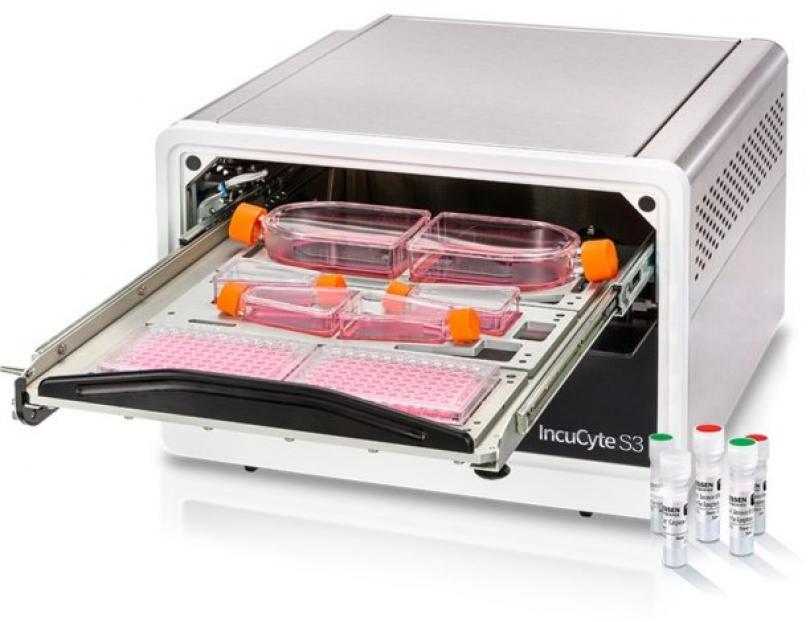
High-throughput Screening |
With a capacity of holding up to 6x 384-well plates the IncyCyte has the potential to screen 2304 wells per experiment. The software comes bundled with analysis tool to measure proliferation and tube formation which can be displayed as plate maps making it easy to see instantly how these parameters change between different wells/treatments. |
|
Wound Healing Assays |
A common method for assessing cell motility is to induce a scratch wound in a confluent monolayer of cultured cells. With a WoundMaker-96 reproducible wounds can be made in 96 well plates and imaged in the IncuCyte. The instrument will automatically measure and report on wound closure. |
|
Proliferation Assays |
The environmental stability that comes from being housed in a tissue culture incubator, along with automated analysis software makes the IncuCyte an attractive tool for performing compound screens. The absence of proliferation is a simple marker for screening drugs that are hoped to induce cell death or senescence. |
|
Apoptosis |
The IncuCyte® Live-Cell Analysis System enables real-time and automated apoptosis assays inside your tissue culture incubator. Two apoptotic pathways assay have been developed to be studied with the Incucyte software module, Caspase-3/7 and Annexin V, and can be studied simultaneously. For more information on reagent and procedure, please go on Incucyte website. |
|
Cytotoxicity |
The IncuCyte® live-cell imaging and analysis system enables you to measure cell death based on cell membrane integrity, in real-time, in automated fashion. |
|
Immune cell proliferation |
You can monitor proliferation and cell-cell clustering interactions without the need to label, in real time and without removing your cultures from the incubator. The incuCyte® software enables accurate quantification of cell clustering over time using phase contrast images. |
|
Tumor Spheroids |
3D spheroid cell culture models are advanced tools to accelerate drug discovery. You can automatically monitor and quantify tumor spheroids information such as growth and health, in real time and inside your tissue culture incubator. |
|
Angiogenesis |
You can follow tube formation/inhibition in real time by acquiring and analyzing images of GFP positive endothelial cells using the integrated Angiogenesis software module. Visualize kinetic plots in microplate views. Export metrics (tube length, tube area, branch points) to calculate EC50/IC50 values. Quantitative Time-course data, measure GFP tube length formation (mm/mm2) |
|
Neurite Dynamics |
You can achieve automated, continuous, non-invasive imaging and measurement of neurite outgrowth with 96-/384-well assays. Quantify neurite length and branch points in order to evaluate neural network stability over time – all inside your cell culture incubator. |
|
Neuronal coculture |
Study and quantify Neurite length (mm/mm2) of labelled neurons in coculture |
|
Chemotaxis |
Quantify cells that migrate through pores and adhere to the bottom-side of a membrane. |