
Welcome to Biological Specimen Preparation (BSP)
High-quality specimens are essential for achieving clear microscopy images. At KGLMF's BSP Laboratory, we offer extensive support to researchers in sample preparation for our imaging equipment, including access to facilities and expert training.
Our Services
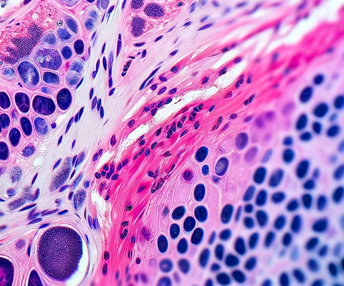 |
HistologyOur in-house histology facility offers a full spectrum of sample preparation services. From tissue embedding, cryo- and microtome sectioning, as well as routine and special histological staining.
|
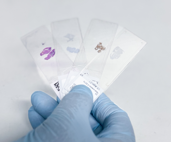 |
Slide ScanningOur slide scanning service provides high-quality brightfield, phase contrast, polarized light, and widefield fluorescence imaging of up to 5 colours..
|
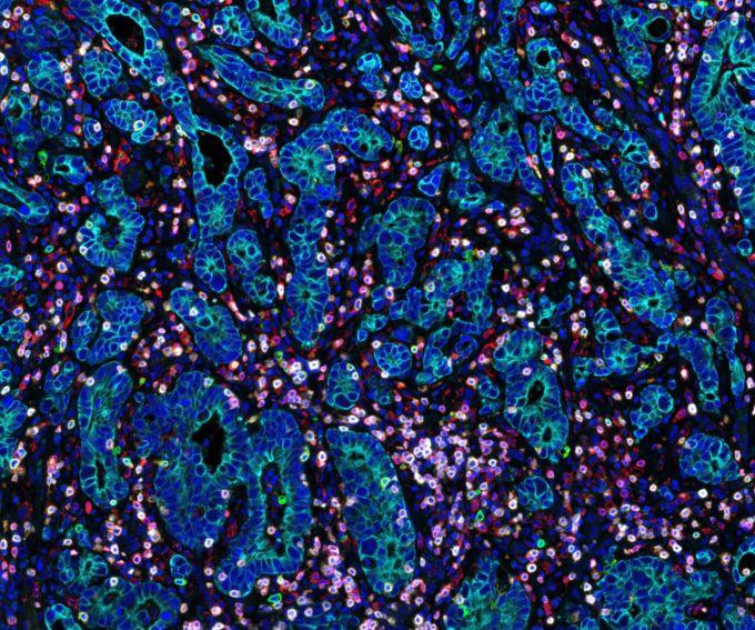 |
MultiplexingGo beyond the limitations of conventional immunofluorescence with our advanced multiplexing service. By utilizing the OPAL-TSA technology, we offer staining of up to 8 markers while preserving the spatial context of your samples.
|