JPK Nanowizard 4 XP with Hybrid stage

jpk-img

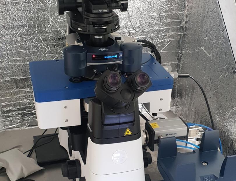
The NanoWizard® 4 XP atomic force microscope is an advanced Atomic Force Microscope (AFM). AFM is a very high-resolution type of Scanning probe microscopy (SPM) which acquires data by “feeling” or “touching” the surface of a sample with a mechanical probe.


This AFM allows the rapid acquisition of a range of sample mechanical properties including elasticity, adhesion and deformation simultaneously with topography and epifluorescence wide field microscopy. The Nanowizard 4 XP features both the Bruker PeakForce Tapping technology and the JPK QI advanced mode.

It can be operated in air and in liquid and is particularly recommended for the study of cells, tissues, biomaterials, single molecules and membranes.

The long range hybrid stage allow cell-cell or cell-substrate force spectroscopy


Hybrid stage Piezo electric scanner: 200 x 200 x 200 µm 3 XYZ scan range

Nanowizard 4 XP head piezo electric scanner: 100 x 100 x 15 µm 3 XYZ scan range

Hybrid stage motorized XY: 20mm x 20mm for large sample tiling, correction of sample tilt

Temperature control: petri dish heater and JPK BioCell II (for coverslips)

Software: V7

probe holder: fixed spring cantilever holder for air and liquid, side view, directdrive.

Deflection detection: IR super luminescent diode λ= 850nm

Topview optical module for opaque samples

Nikon Ti-U inverted microscope(widefield, brightfield & epifluorescence)

High speed Andor Zyla CCD camera, fully integrated

Direct overlay of optical and AFM images


Common Applications

Topographic Imaging: A topographic image is obtained while scanning with the AFM and formed by scale color mapping of the height measured at every pixel of the scan. Advanced scanning modes limits the damage of very soft samples by an accurate control of the force set point (from pN to nN) as well as protecting the probe ensuring consistent, long-term, high-resolution imaging.
Mechanical Properties on soft samples: The AFM supports measurements in PeakForce QNM (Quantitative Nanomechanical Mapping), QI advanced mode and advanced force spectroscopy. QNM and QI allow for the simultaneous acquisition of multiple sample properties including topology, modulus, adhesion and deformation. Both features allow for the capture and analysis of individual force curves at each pixel of the image. These features have been used to image cells, tissues and biomaterials such as hydrogels.
Optical microscopy correlation: The Nanowizard AFM is positioned on a manual inverted widefield epifluorescence microscope (Nikon Ti-U2), allowing for a real time import and registration of optical images with the DirectOverlay™ 2 software. Optical images can be used to guide AFM imaging or force measurements and allow AFM and optical image overlay. 
Single molecules and membranes: The AFM high resolution allows for the imaging and force spectroscopy of Proteins, other biomolecules, and bio molecular assemblies as well as lipid monolayers and bilayers. 
Cell-substrate / Cell-cell spectroscopy: The long z range of the hybrid stage allows for cell-substrate and cell-cell force spectroscopy. A cell, chemically bound to a cantilever, is bought in contact with the sample of interest (molecular layer, single cell, biomaterial…). The user sets the time of contact and a force vs. distance curve is recorded while the cell is separated again, to determine adhesion, viscoelastic parameters, single molecule events as well as tether formation. 
Micro rheology: The microrheology module performs oscillations in z while in contact with the surface, to probe the frequency dependent response of the surface. It is available in the ForceRamp Designer in Advanced Force spectroscopy mode. 


Objective Lenses

MAG N.A Corrections Immersion W.D
4x 0.13 Plan Fluor DL AIR 16.5
10x 0.3 Plan Fluor DLL AIR 1.3
20x 0.5 Plan Fluor DLL AIR 2.1
40x 0.75 Plan Fluor AIR 0.66
60x 1.2 Plan Apo VC WATER 0.31 - 0.28


Light sources

Source Wavelengths
Tungsten Light Transmitted white light
Mercury arc lamp Incident visible light


Filters

Name Excitation Dichroic Emission
DAPI BP 361-389 415 BP 430-490
GFP BP 451-487 497 BP 506-544
YFP BP 489-505 516 BP 524-546
TxRed BP 542-576 585 BP 596-664


Detectors

Name Description
Andor Zyla 5.5 USB3-W Scientific Camera High speed CCD Camera fully integrated in JPK software