Chemicon Advanced Tissue Arrayer

Chemicon image

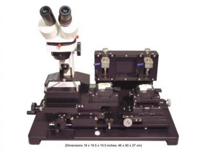
Chemicon Advanced Tissue Arrayer ATA100 allows you to create customized arrays of multiple tissue specimens on a single histologic slide. This instrument allows to selectively remove core tissue samples from paraffin blocks and arrange those tissue samples into an array within a recipient paraffin block. The newly formed tissue array paraffin block is then ready for further histology technique processing. Sections can be stained with the same protocol to avoid experimental variability and technical artefacts. This type of sample preparation is very useful in cancer research, but also anytime a high-throughput morphology or molecular analysis is needed. Tissue microarrays (TMA) in combination with immunohistochemistry has been used to study and validate cancer biomarkers in large numbers of samples in defined cancer patient cohorts.


Material and equipment

Reference slides, donor needle and blocks, recipient needle and blocks, forceps, xylene and array layout table.

Procedures

  • Place reference slide on microscope station.
  • Place donor and recipient needles onto needle holders and place donor and recipient blocks onto block holders.
  • Align donor block and reference slide, set depth of donor needle, set height of recipient block as well as recipient needle.
  • Align recipient needle to centre of recipient block, determine tissue array grid pattern desired and move recipient needle to position A1.
  • Use recipient needle to punch recipient hole, use donor needle to pitch donor core from donor block and deliver donor tissue core into recipient block.
  • Return recipient block under recipient needle and move recipient needle to position B1.
  • Remove needles and blocks, rinse needles with xylene and put them back to the needle boxes and clean up working area after the work finished.