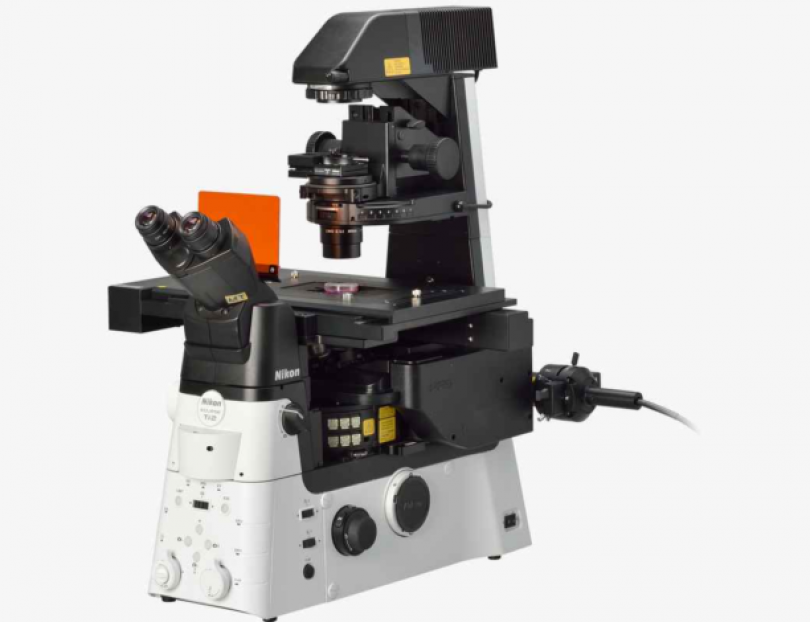
Nikon TIRF/HiLo

Nikon Ti2 image

This microscope combines a total internal fluorescence (TIRF) and highly inclined and laminated optical sheet (HiLo: https://www.nature.com/articles/nmeth1171) modalities in one instrument. The TIRF modality allows user to image molecules at surface of cells and apply super- resolution imaging techniques such as PALM and STORM. HiLo modality offers the possibility of exciting a thin inclined plane within cells or thin layer of cells within few micrometers of a live animal tissue. Automatized variability of angle of HiLo emergence and camera imaging allows for fast sectioning of sample. The system is also supplemented with NIDAQ board and control with external trigger, which combined with the versatile illumination sequence option makes this ideal microscope for complex experiments involving the correct timing of imaging following an external stimulus of cells or animal. Definite focus is perfectly controlled using Nikon PFS system making it ideal for long term live cell or animal imaging. 

Microscope body:
Inverted
Motorised Stage:
Yes
Temp Control:
Fully Enclosed
CO2:
Stage Insert
Software:
NIS Elements & Jobs


Common Applications

Live Cell Imaging: Many biological processes are dynamic events and as such observing living cells is crucial to unravelling and understanding the mechanisms behind them. This requires special considerations when imaging. Firstly, the environmental conditions must be controllable and stable to ensure the cells remain healthy. Secondly care needs to be taken that the act of imaging has as little impact as possible on the event being observed. This microscope has fully enclosed heated chamber and CO2 supply for short or long term live cell imaging.
Cell Migration: This broad term is used to refer to any processes that involve the translation of cells from one location to another. This may include development, wound healing, invasion of cancer cells, immune response. This microscope is equipped with definite focus system, PFS, which is perfect for this type of application. 
TIRF Total Internal Reflection Fluorescence microscopy exploits the unique properties of an induced evanescent wave or field in a limited specimen region immediately adjacent to the interface between two media having different refractive indices. In practice, the most commonly utilized interface in the application of TIRF is the contact area between a cell and a glass coverslip or tissue culture container.
Single Molecule Imaging Single molecule at the surface of a cell using TIRF (N-STORM) modality or within single cross section of cells using HiLo modality. Cooled EMCCD camera is perfect for capturing single molecules at detectable signal to noise. 
Intravital Imaging Intravital imaging is the observation of biological processes in living animals/tissue. It introduces new challenges such as achieving higher penetration depths and dealing with sample mobility. Appreciating that how cells respond in tissue culture has often been shown to be largely different from their response in their true environment makes intravital imaging a crucial step in understanding biological systems. This microscope can do intravital imaging at single molecule level within first few micrometers of an epithelium.


Objective Lenses

MAG N.A CORRECTIONS IMMERSION W.D MISC.
20x 0.75 Plan Apochromat AIR 1 mm DIC/Pol
60x 1.49 Apochromat OIL 0.12 mm DIC/Pol
100x 1.49 Apochromat OIL 0.12 mm DIC/Pol

Note: All lenses are compatible with definite focus PFS. 20 X lens is suitable for dark field microscopy. 60 and 100 x can be used with UV illumination but not recommended. 

Light Sources

Source Wavelengths Output Power
Laser OBIS 405 405 nm 100 mW
Laser OBIS 488 488 nm 100 mW
Laser OBIS 561 561 nm 100 mW
Laser OBIS 640 640 nm 100 mW
Mercury Lamp 400-600 nm  


Filters

 
NAME EXCITATION DICHROIC EMISSION
DAPI WF BP 340 - 380 nm LP 400 nm BP 435 - 485 nm
GFP WF BP 465 - 495 nm LP 505 nm BP 515 - 555 nm
TxRd WF BP 528 - 553 nm LP 565 nm BP 590 - 650 nm


NAME EXCITATION DICHROIC EMISSION
Cy3/Cy5 BP 572 - 621 nm LP 600 nm -
FITC/TRITC

BP 470 - 490 nm

BP 540 - 565 nm

-

BP 503- 530 nm

BP 580 - 620 nm

FITC BP 465 - 595 nm LP 505 nm BP 515- 555 nm


Single Band (405, 488, 561/568, 635 nm) BrightLine and dual (488 +561 nm) BrightLine Sedat laser blocking filters. 

Detector

Model Pixel format Sensor Type Readout Speed (MHz) Description
Andor 897 Ultra 512x512 (16um pixel size) EMCCD 1, 5, 10 and 17 Sensitivity and speed at 56 fps at full 512x512 or 595 fps with 128 x 128 crop mode


Contact People

Dr Renee Whan
A. Prof Renee M Whan

Position: Head of Biomedical Imaging Facility

+61 (2) 9065 1823
+61 409 811 073
Dr Elvis Pandzic
Dr Elvis Pandzic

Position: Senior Lecturer

+61 (2) 9065 2212
+61 409 938 510