Common Applications
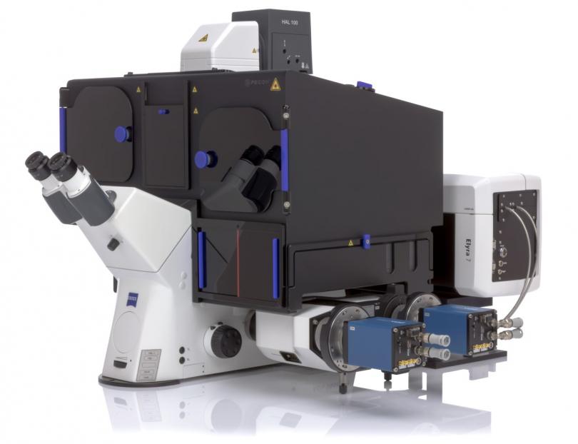
| Lattice SIM | Lattice Structured Illumination Microscopy involves illuminating a fluorescent sample with a ‘lattice’ illumination pattern. A series of images are acquired with the illumination pattern in different orientations, giving spatial information that normally would be lost. The various illumination pattern orientations require 3 rotations and 5 phase shifts for a total of 15 images in order to reconstruct one super-resolution image. Additionally Elyra 7 features ‘Apotome mode’, a faster mode that forgoes the illumination pattern rotations, instead using 5 phase shifts to quickly acquire 2-dimensional image and facilitate optical sectioning. The high speed of the CMOS cameras used in this setup makes this technique ideal for imaging live cells. |
|---|---|
| STORM | Stochastic Optical Reconstruction Microscopy is a super-resolution microscopy technique utilising a TIRF architecture to illuminate the first 100-200 nm of a sample and image single fluorescent molecules. This is achieved by converting fluorophores from a dark to active state using an appropriate buffer solution, before bleaching a short time later. At any time only a very small portion of the fluorophores are active and with each dark-active-bleach cycle more and more of the underlying sample is revealed at up to 10x the resolution of conventional techniques. |
| PALM | Photo-Activated Localisation Microscopy is a super-resolution microscopy technique where the principle of detection and analysis is the same as STORM, but in this case it is not anti-body based labelling and does not require special buffers, but rather requires use of photo-activatable genetically encoded proteins. |
| DNA-PAINT | Similar in principle to STORM and PALM, DNA-based Point Accumulation for Imaging in Nanoscale Topography relies on labels cycling between a fluorescent and dark state to resolve individual molecules. Unlike PALM and STORM, DNA-PAINT achieves cycling through molecular means. Samples are labelled with antibodies conjugated to a single strand of DNA and are treated with a solution of complementary single-stranded DNA conjugated to fluorophores. The continuous binding and unbinding of fluorophore-bound DNA produces a similar cycling when compared to STORM. |


