2015 images of the month

December 2015

Susanne ErdmannSchool of Biotechnology and Biomolecular Sciences (BABS)

December 2015

SE1 is a virus isolated from a hypersaline lake in Antarctica (Deep Lake). It does infect an haloarchaeal strain isolated from the same lake and I am currently getting the genome assembled.

Image by: Susanne ErdmannSchool of Biotechnology and Biomolecular Sciences (BABS)

Microscope/Technique: J1400 TEM, negative staining

November 2015

Hao Wu, School of Chemical Engineering

Hao Wu November 2015

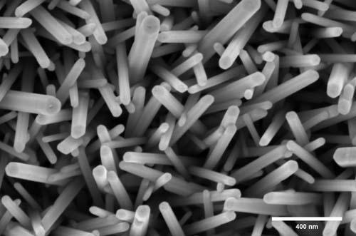
Figure 1 | SEM image of the nanostructures obtained during the growth of ZnO on FTO.

According to the SEM micrograph images the diameter of the ZnO nanorod can be estimated at about 60nm. The entire surface of the FTO glass has been thoroughly coated with ZnO nanorod. Additionally, the width and overall size of the ZnO nanorod seems consistent and not much fluctuation in the dimensions furthermore crediting the success of the chemical bath deposition method to fabricate ZnO nanorod.

My name is Hao Wu (5001090). We are a leading (photo(electro))catalysis research laboratory headed by Professor Rose Amal within the School of Chemical Engineering at the University of New South Wales. The PARTCAT Laboratory evolved from the Centre for Particle and Catalyst Technologies and was part of the ARC Centre of Excellence for Functional Nanomaterials from 2003 until the end of 2013.

Finally, I would like to thank and express my gratitude for Yin Yao and the EMU centre for your guidance during the progress of SEM training. 

October 2015

Lydia Sandiford, School of Chemistry

This is an image of iron/iron oxide core shell nanocrystals for use as MRI contrast agents.

Image by: Lydia Sandiford, School of Chemistry

Microscope/Technique: FEI Tecnai G2 20 TEM

August 2015

Raheleh Pardehkhorram, School of Chemistry

TEM image of gold nanorods which are employed for pathogen detection.

Image by: Raheleh Pardehkhorram, School of Chemistry

Microscope/Technique:FEI Tecnai G2 20 TEM


July 2015

Amanda Wang, Materials Science and Engineering


Where the wild things are - TEM specimens of Ni-Cr plasma sprayed splats on alumina, exhibiting extensive porosity, delamination, and multiple layers of interfacial material.

Image by: Amanda Wang, Materials Science and Engineering

Microscope/Technique: Zeiss Auriga FIB SEM (TEM specimen preparation)