
|
|
The Electron Microscope Unit provides access to advanced electron microscopy and associated sample preparation instrumentation. The units microscopes provide imaging and compositional analysis from the millimeter down to atomic scale, with applications in many fields including advanced manufacturing, medical devices, defense, chemistry, engineering and geology. The Electron Microscope Unit is staffed by eighteen microscopy experts with experience in a wide range of fields. We can also facilitate links with the wider UNSW research community and other advanced analytical equipment located in the Mark Wainwright Analytical Centre. We offer a tailored solution to your problem and can provide fee-for-service or hands-on training for independent access to our microscopes and sample preparation equipment. The Electron Microscope Unit is located on the main UNSW Sydney campus in Kensington with labs in the Chemical Sciences Building (F10), Hilmer Building (E10) and the Science and Engineering Building (E8). (Click for campus map) CONTACT US Please contact us to discuss how we can help you and to obtain a quote.
Dr Simon Hager Industry Applications Scientist 02 9065 2249
|


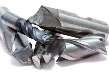 |
Fracture analysis of metals Characterising the fracture mechanism is important for many industries including, defence, aerospace, packaging, transport, 3D printing. Scanning Electron Microscopy is an ideal technique to image a fracture surface and identify the failure mechanism. Understanding the failure mechanism is a critical step in preventing the same failure reoccurring. |
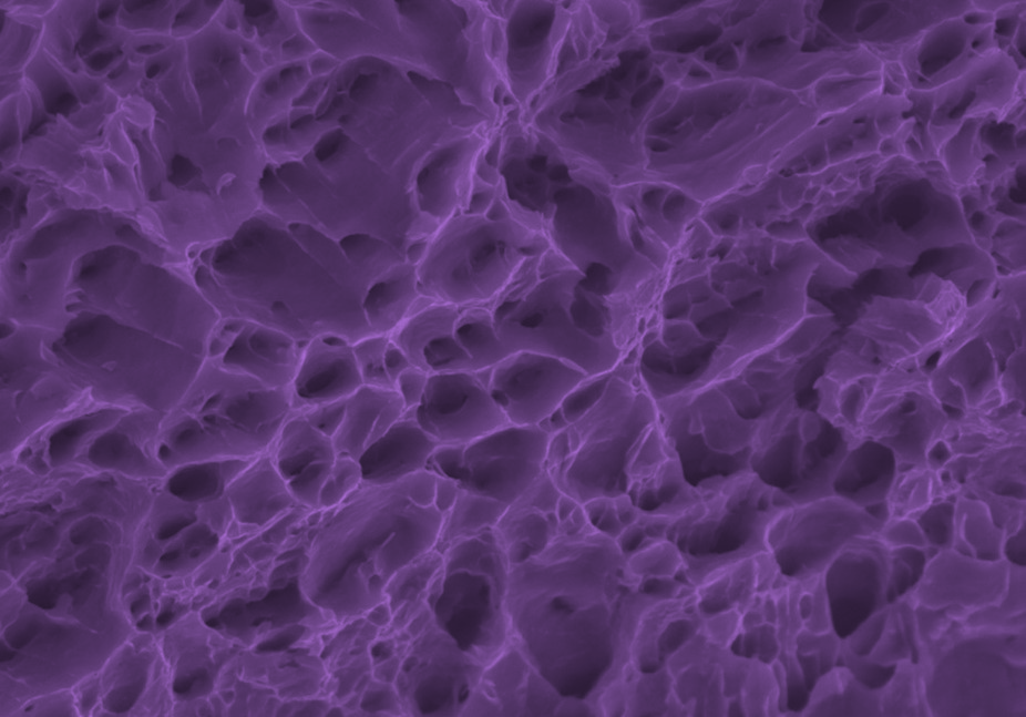 |
SEM image showing the dimple structure (pores) that is characteristic of a ductile fracture, a feature responsible for the failure of a metal packaging sample.
|
|
|
|

|
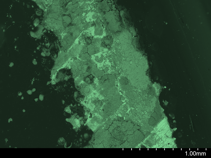
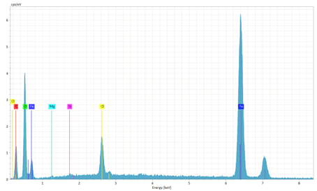
Corrosion identification in Steel Tin platings are used as a coating on steel to prevent rusting and is widely used in food and beverage packaging, electrical parts and wire shielding. Identifying which elements are present in a corroded metal sample can explain why the corrosion has occurred and provide solutions to prevent it recurring which will minimise product waste and increase yield. |
|
Scanning Electron Microscopy has been used to image the location of the corroded steel sample. Energy dispersive spectroscopy can identify which elements are present in the corrosion. In addition to iron and oxygen a significant amount of chloride was detected which is a common cause of corrosion. Identifying that chloride is present in the corrosion products on a tin plate steel indicates why the corrosion has formed
|
|
|
|
|
|
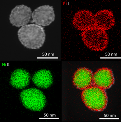
Nanoparticle analysis for catalysts, battery materials and air pollutants The size, morphology and composition of nanoparticles are critical for applications in clean energy, battery materials and environmental contamination. Transmission Electron Microscopy is a versatile tool for characterising nanoparticles from single atoms to several hundred nanometres in size. Transmission Electron Microscopy has been used to characterise the thickness and morphology of a platinum shell over a nickel core. Changes in the thickness and morphology of the Pt shell have been related to an improved yield of hydrogen derived from splitting water molecules. TEM image shows the structure of a nickle and platinum nanoparticle that improves the generation of hydrogen.
|

|
|
|