2019 images of the month

November 2019

Yuan Wang (Helena) - School of Chemistry

NiMoOx particle blossoms

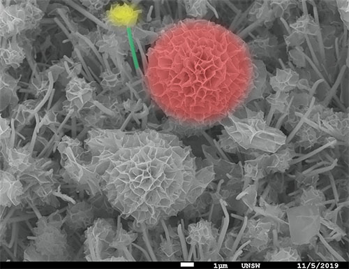
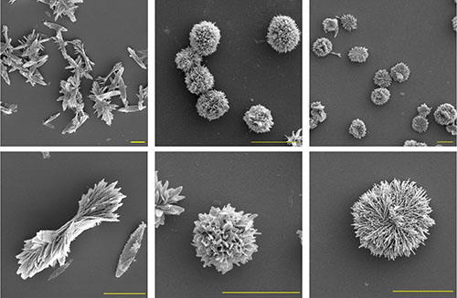
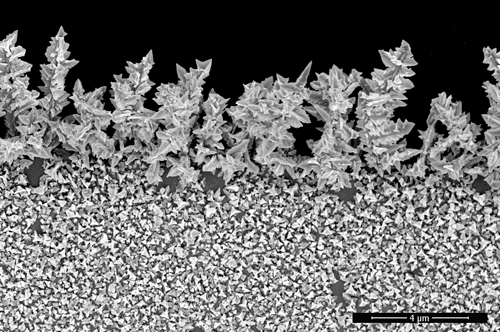
NiMoOx blossom are controllably grown on the backbone of the 3D nickel foam as effective electrocatalyst for water splitting. The stem structure connects the flower to the nickel foam, thereby increasing conductivity and electrochemical surface area.

Image by: Helena Yuan Wang-School of Chemistry

Supervisor: Professor Chuan Zhao

Microscope/Technique: SEM 450

October 2019

Tao Yang - School of Chemical Engineering, Australian Centre for Nanomedicine

Zinc oxide nanoparticles

FE-SEM images illustrate zinc oxide (ZnO) particles with varied morphologies (from left to right: bowtie, flower and nest) synthesized using identical building blocks through a chemical precipitation method. Images were recorded at low (top row) and high (bottom row) resolutions. Scale bar: 5 µm.

Image by: Tao Yang

Supervisor: Dr Rona Chandrawati

Microscope/Technique: NanoSEM 230 and NanoSEM 450

September 2019

Yong Zhao - School of Chemistry

Yong Zhao TEM image

Yong Zhao TEM image 2

This bright field image shows a spontaneous assembly of hexagonal Pd nanosheets with {111} facet exposed on carbon film. four and a half pieces of regular Pd hexagons stacked together by a face-to-face order, which can be clearly discerned in the false-colour strengthened image. the strong electron irradiation induced the formation of nanopores on the surface of the ultrathin nanosheets.

Image by: Dr. Yong Zhao

Supervisor: Prof. Chuan Zhao

Microscope/Technique: cm200 TEM

August 2019

Rokiah Alford and Jonathan Berengut, EMBL Australian Node for Single Molecule Science, School of Medical Sciences

Alford Berengut TEM image

Image by: Rokiah Alford (sample design, synthesis and preparation) & Jonathan Berengut (sample preparation and imaging)

Supervisor: Dr. Lawrence Lee

Microscope/Technique: Tecnai G2 20 TEM/ UA negative stain

July 2019

Reza Shahmiri - School of Materials Science and Engineering

Reza Shahmiri TEM image

This image illustrates a grain of bio-ceramic for dental application. The powder consists of zirconia oxide doped with yttrium. Characteristics of the grain boundary are under investigation with the use of TEM/STEM.

Image by: Reza Shahmiri (with assistance of Richard Webster, Yin Yao and Qiang Zhu)

Supervisor: Dr. Owen Standard

Microscope/Technique:  JEOL f200 TEM

June 2019

Yuwen Xu - School of Materials Science and Engineering

Yuwen Xu TEM image

This HRTEM image shows a very tiny ceramic single crystal nuclei obtained by recrystallisation after long time aging. It exposes {111} and {100} facets and is in the two-dimensional truncated diamond shape with newly grown atomic layers near the corners.

Image by: Yuwen Xu

Supervisors: Prof. Charles C. Sorrell and Dr. Pramod Koshy

Microscope/Technique: f200 TEM

May 2019

Marta Sanchez Miranda - School of Physics

M S Miranda AFM image

3D atomic force microscopy image: InAs nano-membrane with a set of source/drain leads, four hall contacts and an overarching top-gate, insulated by HfO2. this device allows us to gain important insights into the transport properties of these novel InAs structures grown by selective area epitaxy.

Image by: Marta Sanchez Miranda

Supervisor: Prof. Adam Micolich

Microscope/Technique: ICON AFM

April 2019

Hsiang-Sheng (Johnson) Cheng - School of Chemistry

Pt3Sn@Pt core-shell HAADF-STEM

his HAADF-STEM image shows an ordered Pt3Sn@Pt core-shell in cubic shape with {100} facet exposed. The darker part shows the Sn columns while the brighter grids represent the Pt columns in a Au3Cu type ordering structure. 2-3 atomic layers of Pt shell is also observable from the Z contrast.  

Image by: Hsiang-Sheng (Johnson) Chen

Supervisor: Prof. Richard Tilley

Microscope/Technique: JEOL f200 TEM

March 2019

Laicong Qiao - Graduate School of Biomedical Engineering

Gold nanoparticle SEM

We applied electrical technology to control the gold nanoparticle deposition on working electrode. In our experiment, the shape of gold nanoparticle could be successfully controlled (e.g. sphere, flower, dumbbell, popcorn and triangle). In addition, we could also prepare same controlled nanoparticle cluster, such as leaves, seaweed and Christmas tree cluster structures which is shown on the image. This research will provide a reliable surface roughness technology to increase sensitivity of biosensors.

Image by: Laicong Qiao

Supervisor: Dr Guozhen Liu

Microscope/Technique: NanoSEM 450

January 2019

Wilson Handoko - School of Materials Science and Engineering

Iron carbide SEM

SEM micrograph of etched pearlite and micron-sized secondary phase precipitations – iron carbide (Fe3C) on the surface of high carbon steel. One inclusion was located on pearlitic phase surrounded by lamellar structures and another one was occupied along the grain boundary. This microstructure was achieved by ultra-high temperature confocal scanning laser microscope with controlled quenching rate (-60⁰C/min) on originated dual-phase (martensite and retained austensite) high carbon steel.

Image by: Wilson Handoko

Supervisors:  Prof. Veena Sahajwalla and Dr Farshid Pahlevani. 

Microscope/Technique: JEOL 7001f