2016 images of the month


November/December 2016

Jingjing Duan, School of Chemistry

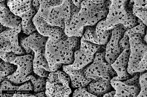
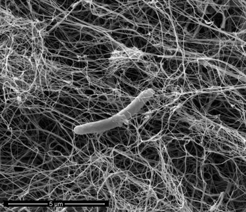
This image is the top view of multi-dimensional nickel sulfide array in situ-grown on nickel foam. The preliminary structure of this nickel sulfide array is the two-dimensional ultra-thin sheets, which constructs to one dimensional wire-like morphology, and further to the three-dimensional array.

Image by: Jingjing Duan, School of Chemistry

Microscope/Technique: FEI NanoSEM 450

Supervisor: A/Prof Chuan Zhao


October 2016

Jon Berengut, Single Molecule Science, School of Medical Sciences

This micrograph depicts DNA origami nanostructures that have been made by designing and synthesising hundreds of single strands and allowing them to self-assemble into precise 3D structures. These blocks have four different-sized cavities for kinetic experiments and an asymmetric hole running through to establish orientation. Inset: A rendering of the design in which sections of double stranded DNA are depicted as cylinders.

Image by:  Jon Berengut, Single Molecule Science, School of Medical Sciences

Instrument used: FEI Tecnai G2 20 TEM

Supervisor: Lawrence Lee


September 2016

Shinyoung NohSchool of Photovoltaic and Renewable Energy Engineering (SPREE)


This image is cross-section view of Ag evaporated on Reactive Ion Etched(RIE) Silica particles on Si wafer. Originally, particles were spherical and closely-packed. RIE deformed particles into smaller ellipsoidal shape with giving distance between the particles. So that evaporated Ag film is patterned. 
 

Image by: Shinyoung NohSchool of Photovoltaic and Renewable Energy Engineering (SPREE)

Microscope/Technique: FEI NanoSEM 450

Supervisor: Dr. Xiaojing Hao


August 2016

Yuan (Helena) Wang, School of Chemical Engineering

The 3D drape porous NiO materials with high specific surface area were fabricated by templating strategy. It is designed as an effective thermal conductive catalyst for CO2 reduction. Comparing to nano particles catalysts, 3D drape porous NiO exhibits extremely high thermal stability, catalytic activity and methane selectivity.

Image by: Yuan (Helena) Wang, School of Chemical Engineering

Microscope/Technique: FEI NanoSEM 230

Supervisors: Scientia Professor Rose Amal and Dr. Hamid Arandiyan


July 2016

Sajjad S. Mofarah, School of Materials Science and Engineering

The image shows hexagonal cerium oxide tubes grown on FTO substrate by the method of anodic electrodeposition. The images taken at very early stages of growing reveal that cerium oxide starts growing in rod shape. However applying potential for a longer time makes them to hollow which finally turns into tube. The tubes with average height of 30 -40 micron is expected to show significant photocatalytic activity.

Image by: Sajjad S. Mofarah, School of Materials Science and Engineering

Microscope/Technique: FEI NanoSEM 450

Supervisor: Prof. C. Sorrell


June 2016

Lucy Gloag, School of Chemistry

The low resolution TEM image shows a superlattice of interlocked Ru hourglass-shaped nanoparticles. The nanoparticles self-assemble on the TEM grid to form an alternating pattern of hexagon and hourglass shapes. The hexagonal base of the hourglass is seen when the nanoparticles are oriented ‘upwards’ and the hourglass shape is seen when the nanoparticles are oriented ’side-on’ to the electron beam.

Image by: Lucy Gloag, School of Chemistry

Microscope/Technique: Phillips cm200 TEM


April/May 2016

Rob RussellSchool of Biological and Biomolecular Sciences

Cellulose fibres are excreted from some bacterial cells and can form a “woven mat” of cellulose with high porosity.

A piece of cellulose pellicle was prepared for SEM using the critical point dryer at the EMU, many thanks to Sigrid Fraser for guidance.

The dried cellulose was imaged using the SEM 230 following platinum coating. The image shows a single Gluconacetobacter xylinus cell whose structure has been maintained by the drying process.

Image by: Rob Russell, School of Biological and Biomolecular Sciences

Instrument used: FEI NanoSEM 230


March 2016

Derrick Lau, School of Medical Sciences

The HIV-1 capsid is a protein cage that protects the fragile genome. A mutant capsid protein has been shown to make uniform spheres (Pornillos et al. 2010). The plasmid were supplied to us to purify. Those proteins do indeed self-assembled into very nice spherical balls!

Methods: Negative staining with Uranyl acetate (2% w/v in water) on formvar coated carbon grids provided by the EMU. 

Image by: Derrick Lau, PhD student, Single Molecule Science, School of Medical Sciences

Instrument used: FEI Tecnai G2 20 TEM


February 2016

Simon Hager, Electron Microscope Unit

This x‐ray map shows the primary sulphide mineralisation in the ore body from the Sunny Corner silver mine which is located approximately 40km to the east of Bathurst. The colours of the individual minerals are;

Purple: pyrite (FeS2),
Green: quartz (SiO2),
Light Blue: galena (PbS),
Dark Blue: sphalerite (ZnS) and
Pink: freibergite (Ag6Cu4Fe2Sb4S13).
 
Image by: Dr. Simon Hager, Technical Officer, Electron Microscope Unit
 
Instrument used: FEI Quanta ESEM


January 2016

Leeora Gubbay-Nemes, School of Biological Earth & Environmental Sciences

This unique animal print pattern is captured in a volcanic rock from Brothers Submarine Volcano (NZ) and is known as a "Ti-magnetite Symplectite". 

This would have formed during eruption of the volcano at a depth of ~2.5km beneath the seafloor. As magma ascended during the eruption a decrease of pressure and loss water content would have caused the breakdown of hydrous amphibole to a more stable mineral phase of Ti-magnetite, forming this animal-like pattern.

Image by: Leeora Gubbay-Nemes, PhD Student, School of Biological Earth & Environmental Sciences, UNSW

Instrument used: EPMA JEOL-8500