
Common Applications
|
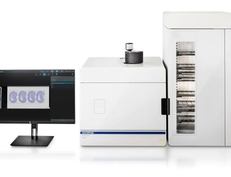
Digitalization of microscope slides |
Tiling and stitching is used to digitalize the image of a tissue slices mounted between a glass slide and a coverslip. The system is accurately calibrated for smooth movements and slide is aligned with detection system. |
|
Brightfield imaging |
Brightfield is used to visualize histologically stained tissue (e.g. H&E, DAB-IHC, Masson’s Trichrome, Picrosirius red...) |
|
Darkfield imaging |
Darkfield is used to enhance the contrast in unstained samples, this trans-illumination widefield technique is only gathering the light scattered by the specimen to form an image. The surrounding, where the beam is not scattered is eliminated and generally appears dark in the image. |
|
Polarization imaging |
Polarization can provide information on color absorption, internal refractive index changes and thickness of the sample. It is using the properties of a birefringent characteristics of the sample to enhance the contrast in the image of a stained or unstained sample. After passing through the sample, the light will pass through an analyzer, grid positioned at exactly 90 degrees from the polarizer, to cancel all polarized wavelengths unaltered by the sample. The subsequent collected light is only able to pass through the analyzer if it interacted with the sample and the degree of interaction is reflected in the final image. |
| Phase contrast imaging | Phase contrast imaging uses an annular ring to block a large portion of the light but will let only a ring-shaped cone of light pass through toward the sample. Where there is no sample, the light will continue its course toward the phase plate, positioned in a way where this untouched light can pass through it without a change. The light interacting with the sample will get diffracted and continues toward the phase plate. The delay of reaching the phase plate creates a contrast in the image. This technique is useful to observe unstained thin samples. |
| Widefield Fluorescent imaging | Widefield Fluorescent microscopy is introducing contrast by using fluorophores into the sample to label structures of interest. LED light source is used to excite fluorophores and the emitted light is then collected on monochromatic camera chip reflecting the change light intensity from one pixel to another. The sample needs to be the thinnest possible as no out-of-focus light is removed from the image. This technique is used to visualize several structures of interest stained by immunofluorescent staining. |
| Virtual z-Stack imaging | The objectives used in this slide scanner have a high numerical aperture. This means that the part of the sample appearing sharp in the collected image will be relatively thin. The slide scanner has capacity to capture z-stacks with variable distance between each plane. This mode enables digitalization of slides thicker than the conventional 4 um tissue sections (up to 300um). |
| Extended Focus Imaging | Extended Focus Imaging (EFI), uses the virtual z-stack function to capture stacks of a thin sample. The software will automatically discard the part of the plane that is not in focus to reconstruct all in-focus regions onto one single image. This is very useful when the tissue sections are thin but uneven. |
| Maximum Intensity Projection Imaging; | Maximum Intensity Projection automatically projects a single plane image composed by the information collected from a z-stack tile scanning. Effectively, the sample is scanned in a z-stack mode and for each pixel only the brightest value across the whole stack is projected in the final image. |