Advanced Search
Pages
BioRep - Procedures
SOPs for Specimen Accrual
Acknowledgement Policy

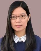
Pearl (Ping) Zhu
Position: Technical Officer
Facility:
Biorepository
Category: General/Technical Staff

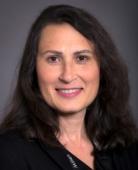
Dr Anusha Hettiaratchi
Position: Manager, UNSW Biorepository
Facility:
Biorepository
Category: General/Technical Staff
BioRep - About Us - Facilities
Specimen storage options
Other services
BioRep - About Us - Aims
Mission Statement
Key Objectives
BioRep - FAQs

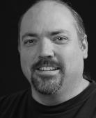
Dr Richard Webster
Position: Research Associate
Facility:
Electron Microscope Unit
Category: Academic Staff
Education
PhD Physics, University of Bristol, UK (2015)
MSci Physics, University of Bristol, UK (2011)
Role
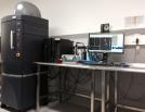
To operate the newly purchased JEOL F200 microscope, design and implement research experiments in collaboration with researchers across the University in the area of advanced transmission electron microscopy, including high-resolution imaging, electron diffraction, energy-dispersive X-ray spectroscopy analysis, and other imaging and analysis modes where applicable.
Research Interests
My interests lie in the application of electron microscopy methods to determine composition, structure and defect nature of semiconductors, mainly III-nitrides. My present research includes the application of electron microscopy to InxGa1-xN nanorods and films for use in LED and transistor devices.
Publications

Shandon Exceisior ES Tissue Processor
Shandon Excelsior ES Tissue Processor is used for automated fixation, dehydration, clearing and wax infiltration of specimens prior to paraffin embedding and sectioning. Subsequent processing like staining and immunostaining can then be performed on processed specimen for pathology diagnosis or research. You can process up to 222 samples at once, across 3 level of organised baskets. The samples are hold into specimen cassettes and each level of organised basket can contain up to 74 cassettes.

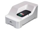
Chemicon Advanced Tissue Arrayer
Chemicon Advanced Tissue Arrayer ATA100 allows you to create customized arrays of multiple tissue specimens on a single histologic slide. This instrument allows to selectively remove core tissue samples from paraffin blocks and arrange those tissue samples into an array within a recipient paraffin block. The newly formed tissue array paraffin block is then ready for further histology technique processing.

Thermo Scientific CryoStar NX70
Thermo Scientific CryoStar NX70 is a high-performance cryostat designed to prepare tissue
sections of frozen samples in medical and research laboratories. Specimen temperature control range is from -50oC to +10oC and blade temperature control range is from -35oC to -5oC. Section thickness can be ranged from 0.5 µm to 500 µm. Fresh or fixed frozen specimens, embedded in OCT, are suitable for cryostat sectioning. Frozen sections could be used for varying staining, such as histology stains, Immunohistochemistry (IFC) and Immunofluorescence (IF) stains.

Leica RM2135 Rotary Microtome
Leica RM2135 rotary microtome is used to cut very thin cross-sections of specimens (usually 4 µm) for routine and research histology studies in the fields of biology and medicine. This instrument is designed for cutting soft and harder paraffin embedded specimens, as long as they are suitable for being cut manually. The sectioning thickness setting range can be from 1 µm to 60 µm. High quality paraffin sections could be used for varying staining, such as Haematoxylin and Eosin stain (H&E), immunohistochemistry (IHC), Immunofluorescent staining (IF) and In situ hybridization (ISH).

EasyGel And SmartClear II
EasyGel is a system designed for speeding the process of producing tissue-gel-hybridization samples. The SmartClear II system is a commercial instrument that allow fast, easy and reliable tissue clearing of tissue-gel-hybridization samples. The system uses stochastic electrotransport technology to remove lipids from tissue-gel-hybridization samples, thereby producing optically transparent samples. Samples that have been optically cleared with the SmartClear II system can be imaged by a number of light microscopy techniques such as confocal and lightsheet.

Leica SP8 DLS
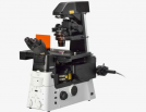
The Leica TCS SP8 DLS (Digital LightSheet) is an inverted laser scanning microscope capable to be used for confocal imaging and light sheet imaging. This microscope is able to image multi-colour, live, fixed or aqueous-based cleared specimens utilising a combinations of 405, 458, 488, 514, 561 and 635 nm excitation lasers through the employment of the AOBS with spectral high quantum efficiency detectors. The advantage of the two combined features (confocal/Lightsheet) on a single equipment allow to perform, with the confocal module, local photo activation or photo bleaching where the resulting dynamics can be capture via lightsheet imaging on thick sample (up to 2 mm thick and 1 cm long).

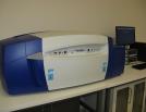
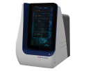
Aperio XT Slide Scanner
The Aperio ScanScope XT scanner is a brightfield microscope that is used to digitalise |

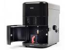
Aperio FL Slide Scanner
The Aperio ScanScope FL scanner is a fluorescence microscope that is used to digitalise traditional fluorescently labelled specimens, mounted on a glass slide, through a high-quality scanning process and produces, able to scan large areas. With 20X magnification you can select from up to 4 fluorescent colors (Blue, Green, red, far-red). All slides must be labelled with DAPI or Hoechst nuclear stain for calibration and focusing purposes.

Leica TCS SP5 WLL STED

TILL Photonics iMic TIRF System

Picoquant Microtime200

Partners
Authorship Guidelines
MWAC adheres to authorship guidelines as set out in the NHMRC Australian Code for the Responsible Conduct of Research as well as the UNSW Research Code of Conduct. Contributions from MWAC staff should be appropriately acknowledged.

Mrs Natasha Kapoor-Kaushik
Position: Technical Staff
Facility:
Electron Microscope Unit
Category: General/Technical Staff
Education
M.Sc (Biotechnology), University of New South Wales, Australia (2008)
B.Sc. (Microbiology), University of Mumbai, India (2004)
Role and Interests
My role is to provide support and training to biological and cryo-electron microscopy users in specimen preparation techniques,
in correlative light electron microscopy and operation of microscopes as well as assist with image acquisition.
Publications

Dr Yin Yao
Position: Technical Officer
Facility:
Electron Microscope Unit
Category: General/Technical Staff
Education
B.E. Material Science and Engineering, University of New South Wales (2010)
Role
Providing training and support for AFM, SEM and Dual Beam FIB.
Research Interests
Powder Metallurgy, Aluminium Alloy, Composites, Ceramics.